Skip to Main Content
  • UHealth |
  • Sylvester Comprehensive Cancer Center

Research

Bascom Palmer Eye Institute
  • Appointments
  • Pay a Bill
  • Referring Physicians
  • International Patients
  • Ways to Give
  • Careers
  • Search
  • Patient Login
  • Find a Doctor
  • Specialties Page 1
    SPECIALTIES
    • Aesthetics
    • Age-Related Macular Degeneration
    • Cataracts
    • Corneal and External Diseases
    • Diabetic Retinopathy
    • General Eye Care
    • Glaucoma
    • LASIK and Laser Vision Correction
    • Neuro-Ophthalmology
    • Ocular Oncology
    • Ophthalmic Plastic and Reconstructive Surgery
    • Pediatric Ophthalmology
    • Retina and Vitreous Diseases
    • Strabismus (Misaligned Eyes)
    • Uveitis
    View all Eye Conditions
    Browse A-Z A B C D E F G H I J K L M N O P Q R S T U V W X Y Z
    SPECIALTIES
    • Aesthetics
    • Age-Related Macular Degeneration
    • Cataracts
    • Corneal and External Diseases
    • Diabetic Retinopathy
    • General Eye Care
    • Glaucoma
    • LASIK and Laser Vision Correction
    • Neuro-Ophthalmology
    • Ocular Oncology
    • Ophthalmic Plastic and Reconstructive Surgery
    • Pediatric Ophthalmology
    • Retina and Vitreous Diseases
    • Strabismus (Misaligned Eyes)
    • Uveitis
    View all Eye Conditions
  • Locations
  • Patients & Families
    Quick links
    • Find a Doctor
    • Specialties
    • Eye Conditions
    • Appointments
    • Locations
    • MyUHealthChart
    • Pay a Bill
    • Insurance Plans
    • Advance Directives
    For Patients & Families
    • Plan Your Visit
    • Preparing for Surgery
    • Eye Emergency Service
    • Rapid Virtual Eye Care
    • Optical Service
    • Low Vision Service
    • International Patients
    • Interpreter Service
    • Medical Records
    • Patient Privacy Rights
    About Bascom Palmer
    • About Bascom Palmer
    • Meet Our Doctors
    • Our Mission
    • Our History
    • Images Magazine
    • News
    • Eye Care Blog
    • Global Impact and Relief Efforts
    • Leadership
    • Careers
    • Contact Us
    • Donate Now

    Need Immediate Eye Care?

    Learn More
    Quick links
    • Find a Doctor
    • Specialties
    • Eye Conditions
    • Appointments
    • Locations
    • MyUHealthChart
    • Pay a Bill
    • Insurance Plans
    • Advance Directives
    For Patients & Families
    • Plan Your Visit
    • Preparing for Surgery
    • Eye Emergency Service
    • Rapid Virtual Eye Care
    • Optical Service
    • Low Vision Service
    • Pharmacy
    • International Patients
    • Interpreter Service
    • Medical Records
    • Patient Privacy Rights
    About Bascom Palmer
    • About Bascom Palmer
    • Meet Our Doctors
    • Our Mission
    • Our History
    • Images Magazine
    • News
    • Eye Care Blog
    • Global Impact and Relief Efforts
    • Leadership
    • Careers
    • Contact Us
    • Donate Now
  • Research
    Clinical Trials
    • About Clinical Studies
    • Understanding Clinical Trials
    • Find a Clinical Trial
    Research
    • About Our Research
    • Research Faculty
    • Research Centers and Laboratories
    • Clinical Research
    • Laboratory Research
    • Research Cores
    Research Resources
    • Research Training
    • Research News

    View our clinical trials

    Start Search
    Clinical Trials
    • About Clinical Studies
    • Understanding Clinical Trials
    • Find a Clinical Trial
    Research
    • About Our Research
    • Research Faculty
    • Research Centers and Laboratories
    • Clinical Research
    • Laboratory Research
    • Research Cores
    Research Resources
    • Research Training
    • Research News
  • Healthcare Professionals
    Referring Physicians
    • Refer a Patient
    • Insurance Plans
    • Ocular Pathology Services
    Education & Training
    • Global Center for Ophthalmic Education
    • Bascom Palmer Learn Portal
    • Continuing Medical Education
    • Ophthalmology Residency Program
    • Fellowship Programs
    • Observership Programs
    • Calendar of Events
    • Grand Rounds
    • Distinguished Lecture Series
    • Medical Student Education
    • Master's in Vision Science (MVSIO)
    • Library Services
    Nursing & Allied Health Professionals
    • Continuing Education for Nurses and Allied Health Professionals
    Alumni
    • Alumni News
    • Alumni Dues

    View our clinical trials

    Translating research breakthroughs into more effective treatments remains one of Bascom Palmer’s highest priorities. Start search
    Referring Physicians
    • Refer a Patient
    • Insurance Plans
    • Ocular Pathology Services
    Education & Training
    • Global Center for Ophthalmic Education
    • Bascom Palmer Learn Portal
    • Continuing Medical Education
    • Fellowship Programs
    • Ophthalmology Residency Program
    • Observership Programs
    • Calendar of Events
    • Grand Rounds
    • Distinguished Lecture Series
    • Medical Student Education
    • Master's in Vision Science (MVSIO)
    • Library Services
    Nursing & Allied Health Professionals
    • Continuing Education for Nurses and Allied Health Professionals
    Alumni
    • Alumni News
    • Alumni Dues
  • Search
Patient Login
  • University of Miami Health System
  • Ranked #1 Eye Hospital in the USA
  • Research
  • Research Centers and Laboratories
  • Neuroinflammation and Ocular Epigenetics Laboratory
  • Research

Neuroinflammation and Ocular Epigenetics Laboratory

Neuroinflammation and Ocular Epigenetics Laboratory

  • Research
  • Lab Members
  • Publications
  • Contact Us

Contact Us

Investigator / Contact Person Dmitry V. Ivanov, Ph.D.
Lab 305-482-4796
Office 305-482-4230
Email divanov@miami.edu

Research

The main research focus of the Neuroinflammation and Ocular Epigenetics Laboratory is to understand the role and mechanisms of sterile inflammation, or innate immune response in the absence of bacteria, viruses, or other microorganisms, in the pathophysiology of retinal disorders, with a specific interest in the contribution of damage-associated molecular patterns (DAMPs) and pattern recognition receptors. Since DAMPs are liberated from necrotic cells mediating the neurotoxic pro-inflammatory response, this laboratory, under the direction of Dmitri Ivanov, Ph.D., also studies the molecular mechanisms of regulated necrosis and its contribution to neuronal damage in retinal neurodegeneration. The second key direction of the laboratory research is the study of epigenetic mechanisms involved in the development and diseases of the eye.

  • Programmed cell death: the role of apoptosis and regulated necrosis in retinal degeneration

    The common forms of vision loss occur because retinal ganglion cells (RGCs) undergo apoptosis and necrosis in the retina. Apoptosis, however, is not as dangerous to the surrounding tissue as necrosis. Apoptotic cell death stimulates the production of anti-inflammatory and neuroprotective factors from immune cells that have internalized apoptotic cells (Fig. 1A). Our studies indicate that therapeutic strategies based on mimicking a systemic increase in levels of apoptotic signals can significantly reduce retinal injury. In contrast, endogenous factors (proteins, RNA, DNA, etc.; known as damage-associated molecular patterns or DAMPs) liberated from necrotic cells mediate cytotoxic, pro-inflammatory responses in retinal tissue (Fig. 1A). We demonstrated that these DAMPs, spilled from necrotic cells, cause inflammation and retinal damage. We also found that suppressing neuronal necrosis in the retina promotes a neuroprotective environment and reduces tissue damage. Thus, in contrast to apoptosis, necrosis can trigger further RGC death and retinal damage. Historically viewed as an entirely accidental and unregulated cellular event, we now know that necrosis, like apoptosis, can be regulated and executed by programmed mechanisms (Fig. 1B). We demonstrated that regulated necrosis contributes to retinal injury through direct loss of RGCs and induction of associated inflammatory responses. Since RGC necrosis is executed via signaling cascades and can be regulated, a better understanding of its molecular mechanisms could lead to discovery of innovative therapeutic strategies for preventing vision loss in patients with retinal neurodegenerative disease. The effectiveness of some of these therapies has already been demonstrated in our published studies.

    Figure 1

    Figure 1. Pathogen–associated molecular patterns (PAMPs) and damage-associated molecular patterns (DAMPs) are responsible for the innate immune response in the tissue. A) Apoptotic cell death stimulates the production of anti-inflammatory and neuroprotective factors from immune cells that have internalized apoptotic cells. Thus, apoptosis is bad news for the cell that triggers this signaling cascade, but good news for the cells that surround it. Meanwhile, cell necrosis is bad news for all neighbors since as a source of sterile inflammation, it triggers a strong cytotoxic pro-inflammatory response in a tissue. B) DAMPs are released only from necrotic cells. Cell necrosis can be accidental or controlled by signaling cascades (regulated/programmed necrosis). C) The products of pathogens (PAMPs) and factors released from necrotic cells (DAMPs) activate the same pattern recognition receptors (PRRs), triggering an inflammatory response. The PRRs include the toll-like receptors (TLRs), NLR receptors, RIG-1-like receptors (RLRs), and the C-type lectin receptors (CLRs).

  • Molecular mechanisms of sterile inflammation in the retina

    Sterile inflammation is a key player in the pathogenesis of many ocular diseases (e.g., ischemic optic neuropathy, glaucoma, optic neuritis, age-related macular degeneration, diabetic retinopathy). But what is the source of sterile inflammation in the retina? By eliminating such source or trigger, degenerative conditions can be improved at the source of the problem, rather than simply concealing symptoms with medications. We have learned over the past decade that after engaging DAMPs (Hsp70, Hmgb1, etc.), pattern recognition receptors (PRRs) such as Tlr4 and Rage activate signaling cascades that trigger inflammation and retinal damage. We also identified new PRR-dependent signaling cascades that mediate retinal damage. We found that the Tlr4/Trif-dependent signaling cascade contributes to retinal damage both directly (through loss of RGCs) and indirectly (through induction of neurotoxic pro-inflammatory responses). The results of our work provide an intellectual foundation for the development of new therapeutic strategies that could reduce neurotoxic, pro-inflammatory responses specifically in the retina without globally suppressing the immune system (Fig.2).

    Figure 2

    Figure 2. The initial stress leads to the accidental RGC necrosis (primary injury) and the release of endogenous factors (EF: e.g., DAMPs [Hsp70, Hmgb1, etc.], glutamate [Glu], ATP, etc.) from necrotic cells. These events trigger two types of mechanisms. A) Inflammatory: The DAMPs act through PRRs in glial cells, causing a toxic pro-inflammatory response (sterile inflammation), and leading to RGC death by apoptosis and regulated necrosis. Regulated necrosis and sterile inflammation, acting as parts of a positive feedback loop, can maintain each other, leading to severe tissue damage (secondary injury). An anti-inflammatory response mediated by apoptotic cell death can stop this vicious cycle. B) Non–inflammatory: EF released from necrotic cells can directly trigger cell death (e.g., Glu excitotoxicity, ATP toxicity, DAMPs such as Hsp70, Hmgb1, etc.) by apoptosis and regulated necrosis without the involvement of pro–inflammatory factors (e.g., TNF, etc.). Regulated necrosis (due to the release of even more EF) reproduces itself (a positive feedback loop), causing significant secondary injury. Meanwhile, since EF are not released from apoptotic cells, this type of programmed cell death prevents subsequent cell death by lowering the levels of EF.

  • The role of the DNA demethylation pathway in retinal development and pathology

    Though much progress has been made in the understanding of signaling cascades driving retinal development and pathology, the role of epigenetic mechanisms remains to be explored as meticulously. To fill this gap, we evaluated the contribution of the DNA methylation and DNA demethylation pathways to retinal development and pathology in recent studies (Fig. 3). We discovered that the promoters (a region of DNA where the transcription of a gene is initiated) of many mutated in retinitis pigmentosa (RP) and related photoreceptor dystrophies genes were highly methylated (hypermethylated) in DNA isolated from fetal retinas and retinal progenitor cells (RPCs; e.g., USH2A, RHO, PRPH2, EYS, AIPL1, CNGB1, IMPG1, IMPG2, NR2E3, PDE6A, PDE6G, PDE6C, PDE6H, RBP3, RP1). The methylation of these promoters was significantly reduced during RPC differentiation into photoreceptors and accompanied by an increased expression of the corresponding genes. It is generally accepted that DNA methylation in promoter regions silences gene expression, while DNA demethylation should occur to allow gene expression. Hence, unsuccessful demethylation of the promoters of the genes listed above during RPC differentiation into photoreceptors may reduce or even eliminate their activity, leading to photoreceptor dystrophies without any mutations in the genomic DNA (Fig. 4). Thus, not only mutations in DNA but also retina-specific epigenetic changes in the DNA may contribute to the pathogenesis of RP and related diseases, indicating the importance of understanding the DNA demethylation pathway during photoreceptor development. The ten–eleven translocation (TET) protein family has a vital role in DNA demethylation and regulates eye development and neurogenesis in various species. Our data and the results of other laboratories indicate that the TET-dependent DNA demethylation pathway controls photoreceptor development. The objectives of this project are to gain a detailed understanding of how the TET-driven DNA demethylation pathway specifies the differentiation of RPCs into photoreceptors, and to investigate how irregularities in its activity lead to photoreceptor death and retinal degeneration.

    Figure 3

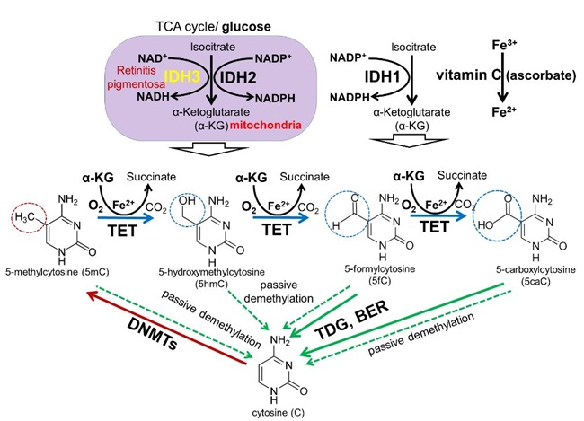
    Figure 3. DNA methylation and demethylation pathways: During development, patterns of methylated cytosines are established by the de novo methyltransferases Dnmt3a/Dnmt3b, and subsequently preserved through cell divisions by Dnmt1. The TET family promotes DNA demethylation by oxidizing 5mC to produce 5hmC, 5fC, and 5caC in DNA. Oxidized derivatives of 5mC inhibit Dnmt1, promoting passive DNA demethylation (dashed lines). 5fC and 5caC are directly excised by thymine DNA glycosylase (TDG) to generate abasic sites triggering base excision repair (BER) pathway activation followed by replacement of the abasic sites with unmodified cytosines.

    Figure 4

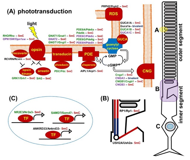
    Figure 4. Promoters of human and mouse genes coding molecular components of the photoreceptor outer segment (phototransduction cascade) and inner segment are mostly hypermethylated in embryonic retinas/RPCs (the corresponding proteins are marked in red). The genes are colored green for rods, purple for cones, and black indicates expression in both rods and cones. (5mC- 5-methylcytosine, gene with hypermethylated promoter; bivalent- bivalent promoter)

  • Epigenetic regulation of retinal development

    Understanding the mechanisms of retinal development is of vital importance in the proposal of therapeutic approaches to restoring visual function via the regeneration of lost retinal neurons. An ideal reparative strategy would be for the retina to heal itself—an inherent characteristic of many species, but not one usually associated with mammals. Adult teleost fish, such as zebrafish, and amphibians have an outstanding self-healing ability to regenerate damaged retinas and restore lost sight. Retinal regeneration in these species relies on two different cell types: Müller glia (MG, teleost fish) and retinal pigment epithelium (RPE, amphibians). MG and RPE undergo a reprogramming process allowing them to generate progenitors for retinal regeneration after injury. Why teleost fish and amphibians have the ability to reprogram MG and RPE after injury, while mammals do not, remains a mystery. We investigated the epigenetic plasticity of RPE and MG to identify possible mechanisms that prevent mammalian RPE and MG from reprogramming and differentiating into retinal neurons. Our published data suggest that epigenetic mechanisms play an essential role in this process.


Quick links
  • Find a Doctor
  • Specialties
  • Eye Conditions
  • Appointments
  • Rapid Virtual Eye Care
  • Locations
  • Insurance Plans
  • Pay a Bill
  • MyUHealthChart
Patients & Families
  • Plan Your Visit
  • Preparing for Surgery
  • Eye Emergency Service
  • Optical Service
  • Low Vision Service
  • International Patients
  • Interpreter Service
  • Medical Records
  • Patient Privacy Rights
  • Price Transparency
  • Florida Health Finder
Medical Professionals
  • Refer a Patient
  • Education & Training
  • Miller School of Medicine
  • Physician News
About Bascom Palmer
  • About Bascom Palmer
  • Meet Our Doctors
  • Our Mission
  • Community Health Needs Assessment
  • Images Magazine
  • News
  • For Media
  • Eye Care Blog
  • Global Impact and Relief Efforts
  • Leadership
  • Careers
  • Contact Us

University of Miami Centennial Logo

Medical Disclaimer | Terms of Use | Privacy Statement | HIPAA Notice of Privacy Practices
© 2025 University of Miami Health System. All rights reserved.