Skip to Main Content
  • UHealth |
  • Sylvester Comprehensive Cancer Center

Research

Bascom Palmer Eye Institute
  • Appointments
  • Pay a Bill
  • Referring Physicians
  • International Patients
  • Ways to Give
  • Careers
  • Search
  • Patient Login
  • Find a Doctor
  • Specialties Page 1
    SPECIALTIES
    • Aesthetics
    • Age-Related Macular Degeneration
    • Cataracts
    • Corneal and External Diseases
    • Diabetic Retinopathy
    • General Eye Care
    • Glaucoma
    • LASIK and Laser Vision Correction
    • Neuro-Ophthalmology
    • Ocular Oncology
    • Ophthalmic Plastic and Reconstructive Surgery
    • Pediatric Ophthalmology
    • Retina and Vitreous Diseases
    • Strabismus (Misaligned Eyes)
    • Uveitis
    View all Eye Conditions
    Browse A-Z A B C D E F G H I J K L M N O P Q R S T U V W X Y Z
    SPECIALTIES
    • Aesthetics
    • Age-Related Macular Degeneration
    • Cataracts
    • Corneal and External Diseases
    • Diabetic Retinopathy
    • General Eye Care
    • Glaucoma
    • LASIK and Laser Vision Correction
    • Neuro-Ophthalmology
    • Ocular Oncology
    • Ophthalmic Plastic and Reconstructive Surgery
    • Pediatric Ophthalmology
    • Retina and Vitreous Diseases
    • Strabismus (Misaligned Eyes)
    • Uveitis
    View all Eye Conditions
  • Locations
  • Patients & Families
    Quick links
    • Find a Doctor
    • Specialties
    • Eye Conditions
    • Appointments
    • Locations
    • MyUHealthChart
    • Pay a Bill
    • Insurance Plans
    • Advance Directives
    For Patients & Families
    • Plan Your Visit
    • Preparing for Surgery
    • Eye Emergency Service
    • Rapid Virtual Eye Care
    • Optical Service
    • Low Vision Service
    • International Patients
    • Interpreter Service
    • Medical Records
    • Patient Privacy Rights
    About Bascom Palmer
    • About Bascom Palmer
    • Meet Our Doctors
    • Our Mission
    • Our History
    • Images Magazine
    • News
    • Eye Care Blog
    • Global Impact and Relief Efforts
    • Leadership
    • Careers
    • Contact Us
    • Donate Now

    Need Immediate Eye Care?

    Learn More
    Quick links
    • Find a Doctor
    • Specialties
    • Eye Conditions
    • Appointments
    • Locations
    • MyUHealthChart
    • Pay a Bill
    • Insurance Plans
    • Advance Directives
    For Patients & Families
    • Plan Your Visit
    • Preparing for Surgery
    • Eye Emergency Service
    • Rapid Virtual Eye Care
    • Optical Service
    • Low Vision Service
    • Pharmacy
    • International Patients
    • Interpreter Service
    • Medical Records
    • Patient Privacy Rights
    About Bascom Palmer
    • About Bascom Palmer
    • Meet Our Doctors
    • Our Mission
    • Our History
    • Images Magazine
    • News
    • Eye Care Blog
    • Global Impact and Relief Efforts
    • Leadership
    • Careers
    • Contact Us
    • Donate Now
  • Research
    Clinical Trials
    • About Clinical Studies
    • Understanding Clinical Trials
    • Find a Clinical Trial
    Research
    • About Our Research
    • Research Faculty
    • Research Centers and Laboratories
    • Clinical Research
    • Laboratory Research
    • Research Cores
    Research Resources
    • Research Training
    • Research News

    View our clinical trials

    Start Search
    Clinical Trials
    • About Clinical Studies
    • Understanding Clinical Trials
    • Find a Clinical Trial
    Research
    • About Our Research
    • Research Faculty
    • Research Centers and Laboratories
    • Clinical Research
    • Laboratory Research
    • Research Cores
    Research Resources
    • Research Training
    • Research News
  • Healthcare Professionals
    Referring Physicians
    • Refer a Patient
    • Insurance Plans
    • Ocular Pathology Services
    Education & Training
    • Global Center for Ophthalmic Education
    • Bascom Palmer Learn Portal
    • Continuing Medical Education
    • Ophthalmology Residency Program
    • Fellowship Programs
    • Observership Programs
    • Calendar of Events
    • Grand Rounds
    • Distinguished Lecture Series
    • Medical Student Education
    • Master's in Vision Science (MVSIO)
    • Library Services
    Nursing & Allied Health Professionals
    • Continuing Education for Nurses and Allied Health Professionals
    Alumni
    • Alumni News
    • Alumni Dues

    View our clinical trials

    Translating research breakthroughs into more effective treatments remains one of Bascom Palmer’s highest priorities. Start search
    Referring Physicians
    • Refer a Patient
    • Insurance Plans
    • Ocular Pathology Services
    Education & Training
    • Global Center for Ophthalmic Education
    • Bascom Palmer Learn Portal
    • Continuing Medical Education
    • Fellowship Programs
    • Ophthalmology Residency Program
    • Observership Programs
    • Calendar of Events
    • Grand Rounds
    • Distinguished Lecture Series
    • Medical Student Education
    • Master's in Vision Science (MVSIO)
    • Library Services
    Nursing & Allied Health Professionals
    • Continuing Education for Nurses and Allied Health Professionals
    Alumni
    • Alumni News
    • Alumni Dues
  • Search
Patient Login
  • University of Miami Health System
  • Ranked #1 Eye Hospital in the USA
  • Research
  • Research Centers and Laboratories
  • Advanced Ophthalmic Imaging Laboratory
  • Research

Advanced Ophthalmic Imaging Laboratory

Advanced Ophthalmic Imaging Laboratory

  • Research
  • Lab Members
  • Publications
  • Contact Us

Contact Us

Investigator / Contact Person Jianhua (Jay) Wang, M.D., Ph.D., M.S.
Office 305-482-5010
Email jwang3@med.miami.edu

Research

Dr. Wang’s research involves optics engineers to develop many prototypes of spectral-domain optical coherence tomography (OCT) devices, including ultra-high-resolution OCT, ultra-long scan depth OCT, dual-channel OCT, magnetomotive OCT, and CMOS camera-based ultra-high-speed OCT. In recent years, Dr. Wang has focused on vascular imaging of the eye and developed the methods and hardware to image microvasculature, microstructure, and microcirculation in the retina and ocular surface. Working with a group of clinicians, he recently focused on microvasculature and microcirculation in the retina as a window of the cerebral vasculature in aging, dementia, and multiple sclerosis.

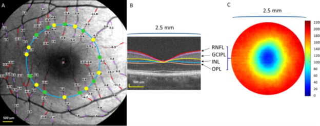
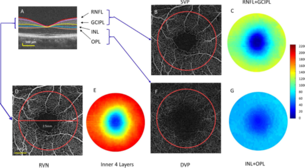
  • Novel approaches for quantitative analysis of microvasculature, microstructure, and microcirculation

    Recently, Dr. Wang contributed significantly to image microvasculature on the ocular surface and retina. A system called functional slit-lamp biomicroscope (FSLB) was developed. This novel system enables easy imaging of the conjunctival microvascular network and small vessel blood flow velocity, which was used to study microvascular response to contact lens wear and changes in dry eye. Working with experts in neuro-ophthalmology, we developed automatic segmentation of retinal microvascular network obtained using Retinal Function Imager (RFI) and optical coherence tomography angiography (OCTA) for studying retinal microvascular changes in multiple sclerosis, AD, diabetics, and cerebral small vessel diseases. In addition, we developed ultra-high-resolution OCT for imaging the retina and adapted Orion segmentation software which can segment seven retinal sub-layers. We visualized focal thickness alterations of intraretinal layers. For the first time, we developed the measurement of retinal tissue perfusion (RTP), volumetric vessel density (VVD), and retinal capillary perfusion (RCP). These measurements have been applied in our research on dementia, multiple sclerosis, and aging. 

  • Retinal microvascular biomarkers for monitoring vascular contribution to dementia

    Easily accessible biomarkers to predict and monitor vascular contribution to cognitive impairment and dementia (VCID) is imperative because accumulating evidence from epidemiology and pathology implicates that microvascular dysfunction is an important contributing factor to age-related dementia. The retinal vessel is regarded as the proxy of small vessels. Retinal and brain vessels share similar anatomic and physiologic features.  Retinal microstructural alterations, such as thinning of the retinal nerve fiber layer and ganglion cell layer, are potential biomarkers of neurodegeneration. In addition, the trending decline of retinal blood flow volume and microvascular network density in cognitive normal controls compared to patients with mild cognitive impairment (MCI), then to Alzheimer’s dementia suggests the presence of vasculopathies in these patients. Dr. Wang’s lab focuses on refining retinal microvascular biomarkers for monitoring vascular contribution VCID. We are studying the ocular changes during normal aging and their relations to cognitive function in patients with cognitive impairment. 

  • Retinal tissue perfusion (RTP)

    Macular blood flow and tissue volume. (A) Retinal blood flow velocity (BFV) was imaged using Retinal Function Imager (RFI). The field of view of 20 degrees was used, and the BFV of the secondary and tertiary branches of the retinal vessels was measured. The arterioles are marked in red and overlaid with the measured blood flow velocities (mm/s). The venules and their respective velocities are marked in pink. A negative value indicates blood flow is moving away from the heart. In this case, the arteriolar flow moved toward the fovea. A positive value indicates blood flow is moving toward the heart. In this case, the vessels are venules. Vessel diameters were calculated at the location where the vessels crossed a circle (2.5 mm) centered on the fovea and the vessel diameters were used to calculate the blood flow in the arterioles (green dots) and venules (yellow dots). The macular blood flow volume of the arterioles was the sum of the blood flow volumes in each arteriole crossing the circle. Similarly, the macular blood flow volume of the venules was the sum of the blood flow volumes in each venule crossing the circle. (B) The intraretinal layers were imaged using UHR-OCT and segmented using Orion software. (C) The thickness of the inner retina including RNFL, GCIPL, INL, and OPL in a disc (2.5 mm). RTP was calculated as the blood flow volume divided by tissue volume (cited from Lin et al. Age-Related Alterations in Retinal Tissue Perfusion and Volumetric Vessel Density. IOVS 2019;60:686).

    RNFL: retinal nerve fiber layer;
    GCIPL: ganglion cell-inner plexiform layer;
    INL: inner nuclear layer;
    OPL: outer plexiform layer.

  • Volumetric vessel density (VVD)

    Tissue volumes and vessel densities of the intraretinal layers. (A) Intraretinal layers were imaged using ultra high-resolution OCT (UHR-OCT) and segmented using the Orion software. (B) The superficial vascular plexus (SVP) of a scan of 3x3 mm was imaged using optical coherence tomography angiography (OCTA) with the analyzed area of a 2.5-mm disc (red circle). (C) The thickness map of the RNFL+GCIPL in a circular area ( 2.5 mm). The VVD of the SVP (VVDs) was the vessel density of the SVP (analyzed as fractal dimension Dbox) divided by the tissue volume of the RNFL and GCIPL in the disc ( 2.5 mm). (D) Retinal vascular network (RVN). (E) The thickness map of the inner retina includes RNFL, GCIPL, INL, and OPL. The VVDr was the vessel density of the RVN (analyzed as fractal dimension Dbox) divided by the tissue volume of the inner retina. (F) Deep vascular plexus (DVP). (G) The thickness map of the INL and OPL in a disc ( 2.5 mm). The VVDd was the vessel density of the DVP (analyzed as fractal dimension Dbox) divided by the tissue volume of the INL and OPL (cited from Lin et al. Age-related alterations in retinal tissue perfusion and volumetric vessel density. IOVS 2019;60:686).

    RNFL: retinal nerve fiber layer;
    GCIPL: ganglion cell-inner plexiform layer;
    INL: inner nuclear layer;
    OPL: outer plexiform layer.

  • Visualization of Focal thickness alteration

    GCIPL Thinning Zone in MS

    Focal thickness alteration of the retinal ganglion cell-inner plexiform layer (GCIPL) in patients with multiple sclerosis (MS). Using different values of the thickness changes on averaged thickness maps, the areas of focal thickness reduction were visualized in MS eyes compared to the control eyes. The most profound thinning zone appeared to locate at nasal 1.98 mm and inferior 0.42 mm from the fovea with a circling zone (diameter f 1 mm), the MS thinning zone of the GCIPL (M zone). The average reduction of the M zone was -7.3 lm in MS eyes (cited from Shi et al. Visual function and disability are associated with focal thickness reduction of the ganglion cell-inner plexiform layer in patients with multiple sclerosis. IOVS 2019;60:1218).


Quick links
  • Find a Doctor
  • Specialties
  • Eye Conditions
  • Appointments
  • Rapid Virtual Eye Care
  • Locations
  • Insurance Plans
  • Pay a Bill
  • MyUHealthChart
Patients & Families
  • Plan Your Visit
  • Preparing for Surgery
  • Eye Emergency Service
  • Optical Service
  • Low Vision Service
  • International Patients
  • Interpreter Service
  • Medical Records
  • Patient Privacy Rights
  • Price Transparency
  • Florida Health Finder
Medical Professionals
  • Refer a Patient
  • Education & Training
  • Miller School of Medicine
  • Physician News
About Bascom Palmer
  • About Bascom Palmer
  • Meet Our Doctors
  • Our Mission
  • Community Health Needs Assessment
  • Images Magazine
  • News
  • For Media
  • Eye Care Blog
  • Global Impact and Relief Efforts
  • Leadership
  • Careers
  • Contact Us

University of Miami Centennial Logo

Medical Disclaimer | Terms of Use | Privacy Statement | HIPAA Notice of Privacy Practices
© 2025 University of Miami Health System. All rights reserved.