
Types of echocardiograms and how to prepare for your appointment
- Learn more about Transthoracic 2D Echocardiography (TTE)
- Learn more about Transesophageal Echocardiography (TEE)
- Learn more about Stress Echocardiography
Transthoracic 2D Echocardiography (TTE)
WHAT IS A TRANSTHORACIC ECHOCARDIOGRAM?
A transthoracic echocardiogram (TTE) is a test that uses ultrasound to create pictures of your heart. With this procedure we can:
- Look at the structure and function of your heart.
- See the size of your heart, and how it contracts.
- See the condition of your heart valves and the blood supply to your heart.
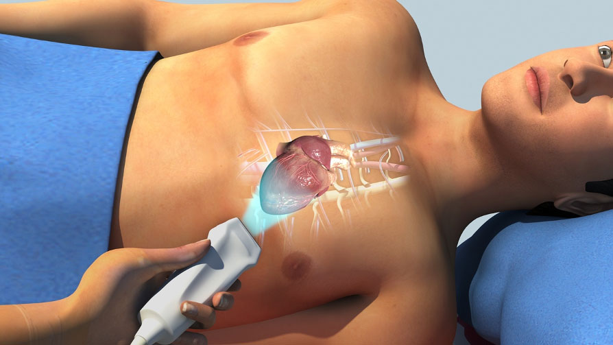
During this test, we place an ultrasound probe on your chest. The high-frequency sound waves from the probe create real-time pictures of the heart. These pictures are displayed on a monitor.
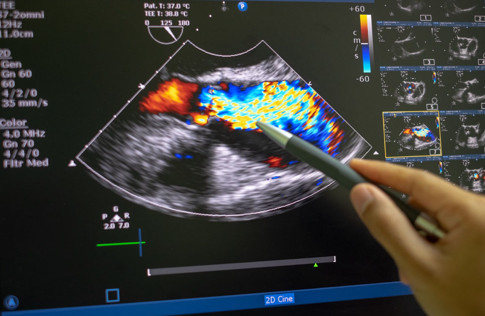
We can also use Doppler technology to look at the speed and direction of blood flow within the heart. TTE is particularly valuable in evaluating:
- the heart’s strength, size, structure, movement, and lining.
- the aorta, a large vessel that takes blood from your heart to the rest of your body.
HOW TO PREPARE FOR YOUR TRANSTHORACIC ECHOCARDIOGRAM
- A TTE does not require special preparation.
- A team member will take you to a private exam room.
- You will need to undress from the waist up.
- You may want to wear a two-piece outfit or loose-fitting clothing.
- A team member will give you a gown to wear.
THE DAY OF THE TEST
- Please arrive at least 30 minutes before your appointment.
- If you are unfamiliar with our location, please plan additional time for travel.
- If you cannot keep your appointment, please call us as soon as possible.
- Please bring your insurance cards and identification with you to the appointment.
- If you have had any prior echocardiograms performed at other facilities, bring a copy of the report to your appointment.
DURING YOUR TEST
- After registration, a team member will take you to a private exam room.
- The team member will give you a gown to put on.
- An ultrasound technologist (the person who does the ultrasound) will place electrodes on your chest to record your heart rhythm and heart rate.
- You will need to lie on your left side on an exam table while the ultrasound technician takes pictures of your heart.
- Some patients may need a contrast agent (a liquid that goes into your body through a vein) to enhance the ultrasound images.
- If this is needed, a team member will place an IV in your hand or arm. We use the IV to administer the contrast agent.
- The typical exam takes about 45 minutes. However, please plan on being in the Cardiology Department for about 90 minutes.
AFTER YOUR TEST
- We store the pictures from the test on digital files.
- The cardiologist reviews the pictures after the test is done.
- We will send a report to your doctor.
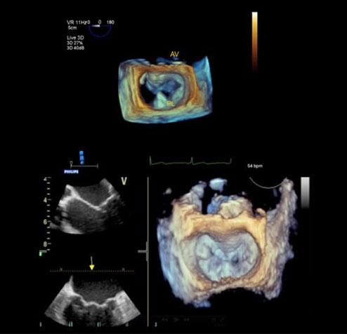
Transesophageal Echocardiography (TEE)
A transesophageal echocardiogram (TEE) is a special type of ultrasound used to look at the structure and function of the heart. A TEE is usually safe.
A TEE is an important tool for taking detailed pictures of your heart, especially when a normal echocardiography may not provide enough information.
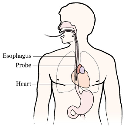
During a TEE the doctor places a small probe into your esophagus (throat). Your esophagus lies close to your heart. The doctor lowers the probe to a spot in your esophagus where it can get clear pictures of your heart’s structures.
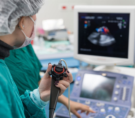
A specialized healthcare team performs the TEE. Before the TEE begins the team will give you medication that relaxes you or puts you to sleep.
A FEW DAYS BEFORE THE PROCEDURE
The TEE nurse will call you a few days before your procedure to:
- Review what you need to do.
- Ask you questions about your medical history.
- Review your medications.
- Tell you which medicines to take and not take the morning of your procedure.
GETTING READY FOR THE PROCEDURE
Before your procedure, please do the following:
- Do not eat or drink for 8 hours before the procedure.
- If you have diabetes, ask your diabetes doctor if you need to adjust your medications.
- Ask someone to drive you home after your procedure. You will be too drowsy to drive.
- Inform your doctor about:
- Any recent bleeding issues.
- Swallowing difficulties.
- Stomach or esophagus (throat) problems.
- Use of a CPAP device.
- Prior issues with anesthesia or medicine allergies
- Before the TEE, your doctor will explain the process and its risks and benefits.
- You’ll need to sign a consent form. This is an ideal time to ask any questions.
DURING THE PROCEDURE
- When you arrive, you will change into a hospital gown.
- A nurse will place an IV into a vein in your arm.
- If you wear dentures, you will remove them before the TEE begins.
- While lying on your left side, the cardiologist will insert the probe into your esophagus.
- You may feel some discomfort, but most people do not feel pain.
- Once the probe reaches the location near the heart, the doctor will take pictures that will help diagnose your condition.
- Although the procedure takes about 30 minutes, expect to be at the hospital for over 2 hours.
AFTER THE PROCEDURE
For your safety and recovery, it is important to know and do the following after your TEE:
- You may be drowsy on and off during the rest of the day.
- Do not drive for the rest of the day.
- Do not operate any machinery.
- It is crucial to have a responsible person stay with you during the first 24 hours after the procedure.
- The medicine that numbs your throat can cause you short-term difficulty swallowing.
- Do not eat or drink for 1 hour after the procedure.
- Once you can drink water without a problem, you may start eating.
Stress Echocardiography
A stress echocardiogram is a test that combines an echocardiogram (an ultrasound of the heart) with a physical stress test. We use this test to determine how your heart functions while exercising.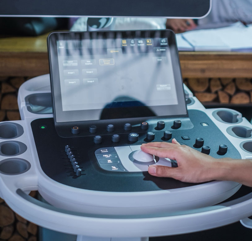
BEFORE THE TEST
- Discuss your medicines with your doctor. You may need to adjust the dose or stop certain medicines before the test.
- We may tell you not to eat certain foods and drinks. Please follow our instructions about fasting or avoiding certain foods and drinks.
- Please do not do activities that need a lot of energy and effort the day before your test.
- On THE DAY OF THE TEST, dress in comfortable, loose clothing that allows you to exercise.
DURING THE TEST
- Before your test starts, a team member places electrodes on your chest to check your heart rate with an electrocardiograph (EKG).
- You will wear a blood pressure cuff to continuously check your blood pressure during the test.
- While you are resting, a team member performs an EKG and echocardiogram.
- For the echocardiogram, you will lie on your left side. At the same time, the ultrasound technician uses an ultrasound wand to capture pictures of your heart.
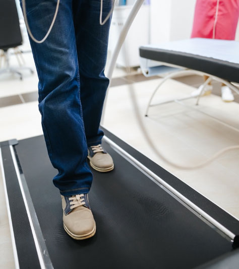
- For the exercise part, you will walk on a treadmill. You will start slowly and then increase your effort.
- You will exercise until you either have symptoms or reach your target heart rate (based on your age and fitness level). It takes about 10 to 15 minutes.
- If you cannot exercise, we will give you a medication called Dobutamine to mimic what your heart does during exercise.
- It is crucial to report any unusual symptoms such as chest pain, shortness of breath, or dizziness to your provider at once.
- After reaching your target heart rate, you will stop exercising and quickly lie down for a repeat echocardiogram.
- It is normal to feel an increase in heart and breathing rate during exercise. You may feel a bit unsteady when stepping off the treadmill.
- The test takes about 1 hour.
AFTER THE TEST
After your blood pressure and heart rate have returned to their normal levels, you can go home.
A cardiologist from our team will review the results of your test. We will share the results with your provider.
Our Mission
The mission of the Echocardiography Laboratory at the University of Miami Health System is to excel in providing state-of-the-art echocardiographic diagnostics, infused with a deep commitment to exceptional patient care. As an academic medical center, we aim to enhance patient outcomes through advanced technology, foster innovation, and educate future leaders in a collaborative, multidisciplinary environment.

Meet the Team
Our Locations

UHealth Tower
1400 NW 12th AvenueSuite 500
Miami, FL 33136

Sylvester Comprehensive Cancer Center
1475 NW 12th Avenue
Ground Floor, Room c905
Miami, FL 33136

Lennar Foundation Medical Center
5555 Ponce De Leon Boulevard
3rd floor
Coral Gables, FL 33146

UHealth at Kendall
8932 SW 97th Avenue
Miami, FL 33176

UHealth at Plantation
8100 SW 10th Street
Suite 2500
Plantation, FL 33324

UHealth at Downtown West Palm Beach
185 Banyan Boulevard
2nd Floor
West Palm Beach, FL 33401
UHealth at Deerfield Beach
1192 E Newport Center Drive
1st floor, STE 100
Deerfield Beach, FL 33442
Questions? We're here to help.
Our appointment specialists are ready to help you find what you need. Contact us today.